Home
/ Arteries Diagram : Human Circulatory System Diagram How It Works Live Science _ Creating a freehand diagram of arteries and veins can be troublesome.
Arteries Diagram : Human Circulatory System Diagram How It Works Live Science _ Creating a freehand diagram of arteries and veins can be troublesome.
Arteries Diagram : Human Circulatory System Diagram How It Works Live Science _ Creating a freehand diagram of arteries and veins can be troublesome.. It ends at the anterior and posterior tibial arteries. This is a congenital defect in which the left pulmonary artery branches off the right pulmonary artery, rather than directly from the pulmonary trunk. Human body artery diagram in detail. More artery diagrams are posted in the following 101 diagramss below. These arteries and their branches supply all parts of the heart muscle with blood.
Ascending aorta, aortic arch, thoracic aorta, and abdominal aorta. Other arteries of the neck. The cardiovascular system consists of the heart, blood vessels, and the approximately 5 liters of blood that the blood vessels transport. The arteries' smaller branches are called arterioles and capillaries. The coronary arteries wrap around the outside of the heart.
Blood Vessel Wikipedia from upload.wikimedia.org These arteries and their branches supply all parts of the heart muscle with blood. Labeled heart diagram showing the heart from anterior. An artery (plural arteries) (from greek ἀρτηρία (artēríā) 'windpipe, artery') is a blood vessel that takes blood away from the heart to one or more parts of the body (tissues, lungs, brain etc.). 5 out of 5 stars. Creating a freehand diagram of arteries and veins can be troublesome. From this trunk, several vessels arise, which go on to supply the neck. The plaque can also burst, leading to a blood clot. All the required arteries and veins on the pancake man.
Most arteries carry oxygenated blood;
More artery diagrams are posted in the following 101 diagramss below. Arteries and arterioles carry oxygenated blood _____ from the heart to the body. The plaque can also burst, leading to a blood clot. Human body artery diagram in detail. It can also help them in getting an overview of artery vs. pulmonary artery sling can be treated surgically. Anatomy and function of the coronary arteries. An artery is an elastic blood vessel that transports blood away from the heart. Finally, the smallest arteries, called arterioles are further branched into small capillaries, where the exchange of all the nutrients, gases and other waste molecules are carried out. The cardiovascular system consists of the heart, blood vessels, and the approximately 5 liters of blood that the blood vessels transport. Arteries are the blood vessels that carry blood away from the heart, where it branches into even smaller vessels. Constricted arteries oppose blood flow, and more pressure is required to push blood. Creating a freehand diagram of arteries and veins can be troublesome.
In this image, you will find right gastric artery, common hepatic artery, celiac trunk, left gastric artery, splenic artery, splenic vein, pancreas, suprarenal vein, renal vein, renal artery, inferior mesenteric vein , gonadal vein, gonadal artery, two alternative position of artery, left colic artery. Other arteries of the neck. Creating a freehand diagram of arteries and veins can be troublesome. 14+ heart arteries diagram labeled. The veins also lack the elastic internal lamina that lies.
Blood Vessel Structure And Function Boundless Anatomy And Physiology from textimgs.s3.amazonaws.com The narrowed arteries are at higher risk for complete blockage from a sudden. Resistance (r) the force opposing blood flow. The first branch of the thyrocervical trunk is the inferior thyroid artery. Arteries of the lower limb thigh leg foot the main artery of the lower limb is femoral artery it is a continuation of the external iliac artery terminal branch of the abdominal aorta the arteries and veins of the leg smartdraw arteries and veins of the leg create healthcare diagrams like this example called arteries and veins of the leg in minutes with smartdraw. Like maps, the various diagrams emphasize different aspects. Arteries of the head and neck diagram art print vintage anatomy art print on tea stained paper dog art dog s wfh office art. Atherosclerosis is a specific type of arteriosclerosis. The neck is supplied by arteries other than the carotids.
Resistance (r) the force opposing blood flow.
This buildup is called plaque. An artery (plural arteries) (from greek ἀρτηρία (artēria) 'windpipe, artery') is a blood vessel that takes blood away from the heart to one or more. A branch of the femoral artery, the popliteal artery branches further to supply blood to the knee, thigh, and calf. Arteries are components of the cardiovascular system. Veins are the blood vessels present throughout the body. The cardiovascular system consists of the heart, blood vessels, and the approximately 5 liters of blood that the blood vessels transport. Finally, the smallest arteries, called arterioles are further branched into small capillaries, where the exchange of all the nutrients, gases and other waste molecules are carried out. Most arteries carry oxygenated blood; Each artery is a muscular tube lined by smooth tissue and has three layers: Over the years, cholesterol plaques can narrow the arteries supplying blood to the heart. From this trunk, several vessels arise, which go on to supply the neck. The plaque can cause your arteries to narrow, blocking blood flow. Arteries of the leg diagram.
This is an excellent human heart diagram which uses different colors to show different parts and also labels a number… Veins are the blood vessels present throughout the body. Anatomy and function of the coronary arteries. Labeled heart diagram showing the heart from anterior. pulmonary artery sling can be treated surgically.
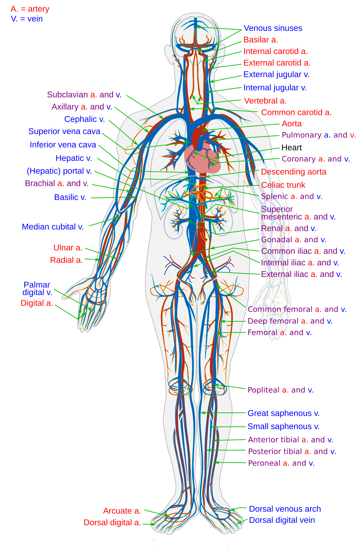
Http Www Wou Edu Lemastm Teaching Bi335 Images 20 20blood 20vessels Pdf from This is the opposite function of veins, which transport blood to the heart. Arteries are the blood vessels that carry blood away from the heart, where it branches into even smaller vessels. In this image, you will find external carotid artery, internal carotid artery, vertebral artery, aorta and arch, pulmonary artery, cardiac artery, thoracic aorta, celiac trunk, superior mesenteric artery, renal artery, gonadal artery, inferior mesenteric artery, common iliac artery, external iliac artery. The aorta branches into a network of smaller arteries that extend throughout the body. Ascending aorta, aortic arch, thoracic aorta, and abdominal aorta. Arteries, veins, and the heart are the main parts of the system. The narrowed arteries are at higher risk for complete blockage from a sudden. An artery (plural arteries) (from greek ἀρτηρία (artēríā) 'windpipe, artery') is a blood vessel that takes blood away from the heart to one or more parts of the body (tissues, lungs, brain etc.).
Arteries, veins, and the heart are the main parts of the system.
An artery (plural arteries) (from greek ἀρτηρία (artēria) 'windpipe, artery') is a blood vessel that takes blood away from the heart to one or more. Two major coronary arteries branch off from the aorta near the point where the aorta and the left ventricle meet. The first branch of the thyrocervical trunk is the inferior thyroid artery. It ends at the anterior and posterior tibial arteries. Constricted arteries oppose blood flow, and more pressure is required to push blood. The right and left subclavian arteries give rise to the thyrocervical trunk. The plaque can also burst, leading to a blood clot. It can also help them in getting an overview of artery vs. The plaque can cause your arteries to narrow, blocking blood flow. John bavosi/science photo library/getty images. The narrowed arteries are at higher risk for complete blockage from a sudden. More artery diagrams are posted in the following 101 diagramss below. The aorta branches into a network of smaller arteries that extend throughout the body.
